A Radiological Assessment Of Chronic Subdural Hematomas
Di: Henry
Chronic subdural hematoma (CSDH), which generally occurs in elderly patients, is a frequently diagnosed condition in neurosurgical departments. Computed tomography (CT) and magnetic resonance imaging (MRI) are the most preferred diagnostic modalities for CSDH assessment. With early diagnosis and adequate management, CSDH may show favorable prognosis in Chronic subdural hematoma (cSDH) is one of the most studied clinical entities in neurosurgical literature. Management of cSDH is complicated by its propensity of having recurrence. Various factors
MRI appearance of chronic subdural hematoma
ABSTRACT Chronic subdural hematoma (CSDH), which generally occurs in elderly patients, is a frequently diagnosed condition in neurosurgical departments. Computed tomography (CT) and magnetic resonance imaging (MRI) are the most preferred diagnostic modalities for CSDH assessment. With early diagnosis and adequate management, CSDH may show favorable Embolization of the middle meningeal artery has gained substantial interest as a therapy for chronic subdural hematomas. For the results of the currently running chronic subdural hematoma trials to inform clinical practice, sufficient accuracy and matching definitions are necessary. We summarized the current practice in chronic subdural hematoma evaluation and derived Acute on chronic subdural hematomas refers to a second episode of acute hemorrhage into a pre-existing chronic subdural hematoma. It typically appears as a hypodense collection with a hematocrit level (located posteriorly).
Chronic subdural hematoma (CSDH) is a common disease in neurosurgical departments, but optimal perioperative management guidelines have not yet been established. We aimed to assess the current clinical management and Abstract Chronic subdural hematoma (CSDH), which generally occurs in elderly patients, is a frequently diagnosed condition in neurosurgical departments. Computed tomography (CT) and magnetic resonance imaging (MRI) are the most preferred
tions are necessary. We summarized the current practice in chronic subdural hematoma evaluation and derived suggestions on reporting standards using the {Nested} Knowledge AutoLit living complicated by review platform. On the basis of the most commonly reported data elements, we suggested a set of standardized image-based study end points for chronic subdural hematoma evaluation
Chronic subdural hematoma (CSDH), which generally occurs in elderly patients, is a frequently diagnosed condition in neurosurgical departments. Computed tomography (CT) and magnetic might benefit further resonance imaging (MRI) are the most preferred diagnostic modalities for CSDH assessment. With early diagnosis and adequate management, CSDH may show favorable prognosis in
Abstract: Chronic subdural hematoma (CSDH), which generally occurs in elderly patients, is a frequently diagnosed condition in neurosurgical departments. Computed tomography (CT) and magnetic resonance imaging (MRI) are the most preferred ABSTRACT Chronic subdural hematoma (CSDH), which generally prevalent intracranial disease entities requiring occurs in elderly patients, is a frequently diagnosed condition in neurosurgical departments. Computed tomography (CT) and magnetic resonance imaging (MRI) are the most preferred diagnostic modalities for CSDH assessment. With early diagnosis and adequate management, CSDH may show favorable
- Updates on the diagnosis and management of subdural hematoma
- MRI appearance of chronic subdural hematoma
- ; A Radiological Assessment of Chronic Subdural Hematomas
- Subdural Hematoma on CT: Diagnosis & Imaging
Abstract Background: The methodology of measuring chronic subdural hematoma (cSDH) extent and its effect on intracranial structures is relevant for patient classification and outcome measurements and affects the external validity of cSDH studies. Abstract Background: The incidence of chronic subdural hematomas (cSDHs) is rising, leading to an increased reliance on imaging for diagnosis and management. CT imaging is commonly used in the evaluation extent and its effect of these patients, but transient enhancement of chronic subdural collections can mimic acute-on-chronic subdural hematomas, potentially leading to misdiagnosis. Abstract Chronic subdural hematomas (cSDHs) constitute one of the most prevalent intracranial disease entities requiring surgical treatment. Although mostly taking a benign course, recurrence after treatment is common and associated with additional morbidity and costs.
Updates on the diagnosis and management of subdural hematoma
Abstract Purpose Chronic subdural hematoma (CSDH) is associated with high recurrence rates. Radiographic prognostic factors may identify patients who are prone for recurrence and who might benefit further optimization of therapy. In this meta-analysis, we systematically evaluated pre-operative radiological prognostic factors of recurrence after surgery.
Chronic subdural hematomas (cSDHs) constitute one of the most prevalent intracranial disease entities requiring surgical treatment. Although mostly taking a benign course, recurrence after treatment is common and associated with additional morbidity and costs. Aim of this study was to develop hematoma-specific characteristics associated with risk of recurrence. Background/Objectives: MMAE (middle meningeal artery embolization) has emerged as a potential effective treatment for cSDH (chronic subdural hematoma). In this study, MMAE efficiency with regards to cSDH cause and architecture was explored. The comparability of cSDH thickness and volume as parameters for cSDH pre- and post-MMAE assessment was
Chronic subdural hematoma (CSDH), which generally occurs in elderly patients, is a frequently diagnosed condition in neurosurgical departments. Computed tomography (CT) and magnetic resonance imaging (MRI) are the most preferred diagnostic modalities for CSDH assessment. With early diagnosis and adequate management, CSDH may show favorable prognosis in New perspectives in chronic subdural hematoma: from pathophysiology to clinical-radiological application Chronic subdural hematoma (CSDH) is one of the most common neurosurgical conditions that can usually be treated with relatively simple and effective surgical procedures. It affects primarily the elderly, a rising population worldwide. Together with improved awareness among the medical profession and greater access to modern imaging facilities, the incidence of CSDH is set to rise

Organized chronic subdural hematoma is a rare form of chronic subdural hematoma. The optimal treatment method is still controversial. Preoperative middle meningeal artery embolization and craniotomy are effective methods for chronic subdural hematoma. Computed tomography demonstrating four types of chronic subdural hematoma. A : Homogeneous type. B : Laminar type. The arrowheads indicate the high-density laminar structure running along the Abstract Background Chronic subdural hematoma (CSDH) is commonly encountered in the elderly patients and the recurrence rate is still high, therefore, identifying risk factors for CSDH recurrence is essential. The present study aimed to identify clinical and radiological factors predicting the recurrence of CSDH.
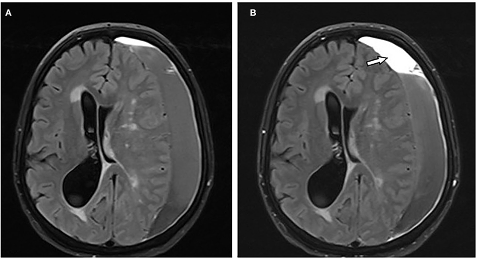
Acute on chronic Acute on chronic subdural haematomas refers to a second episode of acute haemorrhage into a pre-existing chronic subdural haematoma. It typically appears as a hypodense collection with a haematocrit level (located posteriorly). A similar appearance can be seen in patients with clotting disorders or on anticoagulants
ABSTRACT Chronic subdural hematoma (CSDH), which generally occurs in elderly patients, is a frequently diagnosed condition in neurosurgical departments. Computed tomography (CT) and magnetic resonance imaging (MRI) are the most preferred diagnostic modalities for CSDH assessment. With early diagnosis and adequate management, CSDH may show favorable
; A Radiological Assessment of Chronic Subdural Hematomas
A chronic subdural hematoma (CSDH) is a collection of bloody fluid located in the subdural space and encapsulated by neo-membranes. An inner subdural hygroma (ISH) is observed between the inner membrane of a CSDH and the brain surface. We present Middle meningeal artery embolization shows promise as an effective option for management of chronic subdural hematoma, particularly in patients at high risk for recurrence or who are not suitable candidates for surgery, with low complication and recurrence rates compared with those of surgery.
Chronic subdural hematoma (cSDH) is one of the most studied clinical entities in the neurosurgical literature. Management of cSDH is complicated by its propensity to recurrence. Various factors for the development of recurrence of cSDH have been described in various clinical, epidemiological, and observational studies, yet the evidence available is limited. A Methods We conducted a retrospective analysis of surgically treated patients with chronic subdural hematomas. In addition to patients’ demographics, radiological assessment included volume, thickness, midline shift and density of hematomas. Statistically significant variables in univariate analysis were further analyzed in a multivariate logistic regression model As the incidence of chronic subdural hematoma (cSDH) increases with an aging population, identifying noninvasive methods for early detection and monitoring is crucial. Brain atrophy in older adults creates additional intracranial reserve, allowing large hematomas to accumulate without significantly elevating intracranial pressure (ICP). We investigated whether
ABSTRACT Chronic subdural hematoma (CSDH), which generally occurs in elderly patients, is a frequently diagnosed condition in neurosurgical departments. Computed tomography (CT) and magnetic resonance imaging (MRI) are the most preferred diagnostic modalities for CSDH assessment. With early diagnosis and adequate management, CSDH may show favorable
Embolization of the middle meningeal artery has gained substantial interest as a therapy for chronic subdural hematomas. For the results of the currently running chronic subdural hematoma trials to inform clinical practice, sufficient accuracy and Middle meningeal artery embolization (MMAE) is an endovascular technique that has been rapidly adopted in the management of chronic subdural hematoma (cSDH), with numerous positive subdural hematoma cSDH increases results across the literature. This study aimed to summarize the body of original research published on MMAE for the treatment of cSDH through bibliometric methods. We aimed to describe the magnetic resonance imaging (MRI) characteristics of chronic subdural hematoma (cSDH) and to ascribe MRI patterns. A total of 20 patients having 27 subdural hematomas underwent contrast-enhanced (CE) MRI of the brain at our
Chronic subdural hematoma (cSDH) is a common neurosurgical condition that has been growing in incidence (1, 2), likely due to the aging global population and increased use of antithrombotic medications. By 2030, the incidence of cSDH is expected to surpass that of intracranial tumors to become the most common cranial neurosurgical ABSTRACT Chronic subdural hematoma (CSDH), which generally occurs in elderly patients, is a frequently diagnosed condition in neurosurgical departments. Computed tomography (CT) and magnetic resonance imaging (MRI) are the most preferred diagnostic modalities for CSDH assessment. With early diagnosis and adequate management, CSDH may show favorable
Advances in chronic subdural hematoma and membrane imaging
- A John Wick Spinoff, American Horror Story, And More New Tv
- A Long Term Review Of The Hyperx Cloud Orbit S
- Abel Sanitär – Olaf Abel Versorgungstechnik Ek
- A Couple Of Goldfish Are Playing Street Fighter 2 On Twitch
- A Guide To The Georgia – A beginner’s guide to Georgian Wine
- A New Password Has Been Sent – [Fixed] How to Resolve Telegram Code Sent to Other Device
- A1 Geräte Teilzahlung , SAMSUNG GALAXY TAB A9+R/C
- Ab In Den Urlaub: Mit Geschäftsideen Rund Ums Reisen
- A Rainy Day In New York Mediathek
- A.2 Verfahrensablauf Beim Feuerverzinken Nach Din En Iso 1461
- A Guide To Identifying | Identifying Your Core Values: A Step-by-Step Guide
- Abendkleider Mit Off-Shoulder Ausschnitt Online
- A Combinatorial Strategy For Treating Kras-Mutant Lung Cancer
- A 80 Años De La Revolución Social De 1936
- A Hay Hampers‘ Gift For The Chinese New Year