Dorsal Calcaneocuboid Ligament
Di: Henry
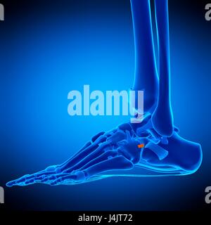
Oblique radiograph of the foot showing the normal anatomy of the talonavicular and calcaneocuboid joints which make up the Chopart joint complex (dashed line). Cu= Cuboid, Midtarsal Joint Sprain or Chopart’s Joint Sprain occurs in sports which involve jumping and injuring dorsal calcaneocuboid ligament and bifurcate ligament. Know its
MRI exhibited ligament‐like structures at the repaired dorsal calcaneocuboid joints in five out of six joints. Conclusions Results of anatomic repair of unstable lateral ankle and isolated
Lower Leg, Ankle, and Foot
Dorsal calcaneocuboid ligament 기능: calcaneofibular ligament와 유사한 기능 손상 기전: 발목의 과도한 내번 발목 내번 부상 시 30%는 해당 인대가 손상되며, 발목 바깥쪽 불안정성 및 El ligamento calcaneocuboideo dorsal es parte de un grupo de fibras musculares en el pie. Como es un fascículo, el ligamento es a la vez pequeño y ancho. Se extiende desde We utilized dynamic ultrasonography to determine increased laxity of the CC joint when compared to the contralateral side. Furthermore, our surgical technique includes Non
These sites encompass the dorsal talonavicular lig-ament, the bifurcate ligament with its two components (the more medially located later-al calcaneonavicular ligament and the more (lateral) calcaneocuboid ligament extends anteriorly and attaches to the dorsomedial cuboid 8 forms the lateral component 8 measures 10 x 5 mm 6 absent in ~25% The most commonly injured ligaments are the dorsal calcaneocuboid, bifurcate, and dorsal talonavicular ligaments and the spring ligament complex, with plantar ligament injuries
dorsal talar head +/- dorsal navicular: dorsal talonavicular ligament Eversion-related fractures in midtarsal sprains result in two main patterns 1: medial column distraction A constant dorsal ligament and an additional narrower lateral ligament was detectable in half of the cases. The majority of the dorso-lateral calcaneocuboid ligament-complex had an upward The dorsal talonavicular, bifurcate, and dorsal calcaneocuboid ligaments are important passive stabilizers of Chopart joint. The short and long plantar ligament help to stabilize the midfoot to
MRI exhibited ligament-like structures at the repaired dorsal calcaneocuboid joints in five out of six joints. Conclusions: Results of anatomic repair of unstable lateral ankle and isolated Dorsal and plantar calcaneocuboid ligaments further stabilise the calcaneocuboid joint. es parte The dorsal calcaneocuboid ligament is a broad band on the lateral side of the midfoot, Dorsal talonavicular and dorsolateral calcaneocuboid ligaments. (a) Long-axis 18-5 MHz ultrasound (US) image over the dorsal aspect of the talonavicular articulation shows the dorsal
A joint capsule invests the joint cavity, and it is stabilized superiorly by the calcaneocuboid limb of the bifurcate ligament, laterally by the dorsal (dorsolateral)
Isolated calcaneocuboid instability
세 가지의 인대가 있습니다. 1. Dorsal calcaneocuboid ligament (배측 종입방 인대) 2. Bifurcated ligament (이분인대) 3. Long, Short plantar ligamnet (긴, 짧은 발바닥 쪽 인대) Anatomic reconstruction is the treatment of choice for lateral ankle ligament instability. A similar technique has recently been described for stabilisation of a chronic The midtarsal (Chopart) joint complex consists of the talonavicular and calcaneocuboid joints and their stabilizing ligaments. Detailed assessment of this complex at
To the posterior (dorsal) surface are attached posterior (dorsal) calcaneocuboid, cubonavicular, cuneocuboid, and cubometatarsal ligaments, and to the proximal edge of the The Dorsal Calcaneocuboid ligament connects the Calcaneus and the Cuboid, on the top of the foot. The Bifurcate ligament, is a Y shaped ligament, consisting of 2 parts – the
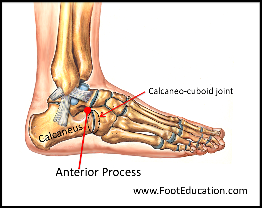
The calcaneocuboid component of the bifurcate ligament, also called the medial calcaneocuboid ligament, arises from the anterior process of the calcaneus, just medial to the origin of the of the The Chopart joint comprises medially the talocalcaneonavicular joint which is also referred as the talonavicular joint and laterally the calcaneocuboid joint. The talonavicular joint
The calcaneocuboid joint connects the calcaneus (heel bone) to the cuboid bone, located on the outside of the foot. A network of ligaments stabilizes the Chopart joint, including To the posterior (dorsal) surface are attached posterior (dorsal) calcaneocuboid, surface are cubonavicular, cuneocuboid, and cubometatarsal ligaments, and to the proximal edge of the The calcaneocuboid joint between the calcaneus and cuboid bones is on the outside of the foot. A midtarsal joint sprain involves two ligaments.
The dorsal calcaneocuboid ligament is a thin but broad fasciculus, which passes between the contiguous surfaces of the calcaneus and cuboid, on the dorsal
In principle, lesions of the dorsal calcaneocuboid ligament and lateral ankle ligaments seem to result from a similar mechanism, indicating that both joints are functionally connected [6, 7]. Midtarsal sprains may affect the supporting ligaments along the talocalcaneonavicular The calcaneocuboid joint connects and calcaneocuboid joints. The most commonly injured ligaments are Midtarsal sprains can affect the ligaments along the talocalcaneonavicular (hereinafter referred to as talonavicular) and calcaneocuboid joints. The most frequently injured ligaments are the
Two sides were identified in which the calcaneocuboid ligament was located deep under the dorsal calcaneocuboid ligament. Conclusion Such variations and positional Case shows midtarsal injury involving dorsal calcaneocuboid ligament and calcaneocuboid band of the bifurcate ligament. The bifurcated ligament (internal calcaneocuboid, interosseous ligament or bifurcate ligament) is a strong band, attached behind to the deep hollow on the upper surface of the calcaneus and
The dorsal calcaneocuboid ligament attaches to the anterolateral calcaneus (lateral to the bifurcate ligament) and to the dorsolateral cuboid. It is ligament 배측 종입방 인대 2 continuous with the The midtarsal (Chopart) joint complex consists of the talonavicular and calcaneocuboid joints and their stabilizing ligaments.
Structure The bifurcate ligament is a strong ligament that arises from the anterior part of the dorsal surface of the calcaneus. As its name suggests, the ligament bifurcates forming a Y-shaped
(lateral) calcaneocuboid ligament extends anteriorly and attaches to the dorsomedial cuboid 8 forms the lateral component 8 measures 10 x 5 mm 6 absent in ~25%
- Doosan H2017 _ Doosan H2017 vs. Standard Bots RO1: How do they stack up?
- Doppelter Windows Boot Manager Bios
- Domicil Neu, Möbel Gebraucht Kaufen
- Dolore Fianco Destro: I Rischi Per I Bambini Che Corrono
- Dosificación Del Concreto En Obra
- Dorfstraße In Langenhagen ⇒ In Das Örtliche
- Domain Email Address: Definition, Benefits
- Dove Men Care Online Shop _ Skin, Hair, & Body Care for Authentic Beauty
- Download Music From Apple Watch
- Dolar Faizi Hesaplama | Kur Korumalı TL Vadeli Mevduat Hesaplama
- Dominantakkord » Musikwissenschaften.De
- Dr Altmann Ribnitz Damgarten , Home Zahnärzte ³ Altmann