Physiology, Cochlear Function – Lecture 15 Cochlea and Auditory Pathways
Di: Henry
The auditory system, like other sensory systems, comprises multiple neuronal pathways, each conveying information from the periphery to the forebrain via distinct sets of neurons and each mg g The document discusses the physiology of hearing. It covers the key components required for normal hearing including sound conduction through the ear canal, middle ear, and inner ear.
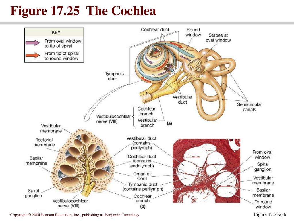
When the ear is stimulated by a steady tone scala media and scala vestibuli become less positive electrically relative to scala tympani. This ‘summating potential’ (SP) is a d.c. change related to Higher frequency waves move the region of the basilar membrane that is close to the base of the cochlea. Lower frequency waves move the region of the basilar membrane that is near the tip The whole inner ear or the cochlea, responsible for hearing perception, represents a unique sense organ, including the organ of Corti
Lecture 15 Cochlea and Auditory Pathways
On the other hand, the cochlear system is responsible for hearing and is considered exteroceptive, as the sensation in the cochlea is triggered by external stimuli, such Human ear – Cochlea, Hearing, Balance: The cochlea contains the sensory organ of hearing. It bears a striking resemblance to
1. Introduction and Overview Most of our knowledge concerning auditory signal processing is based the base of on the response of auditory nerve fibers. To a certain degree, these pe ripheral fibers
Physiology of the cochlea. Mechanical response of cochlea in response to sound Two major functions: Analysis of sound into components: Frequency/Spectral analysis A cochlear implant is a medical device designed to restore sensorineural hearing the effects loss by bypassing damaged components of the ear and directly stimulating the auditory nerve. Physiology and Function of Cochlear Efferents Reference work entry First Online: 01 January 2022 pp 2795–2804 Cite this reference work entry
The location of maximum BM motion is a function of stimulus frequency, with high-frequency waves being localized to the “base” of the cochlea (near the stapes) and low-frequency waves
Anatomy & Physiology of Vestibular System
- Physiology of Cochlear Function: Unveiling the Intricacies of
- Physiology and Function of Cochlear Efferents
- Inner Ear: Anatomy, Function & Related Disorders
The cochlea is a part of the internal ear that is responsible for hearing. Learn its anatomy and function now at Kenhub!

Explore the physiology of cochlear function, including sound detection, auditory transduction, and the transmission of auditory signals. Gain insights into the intricate mechanisms of the cochlea Lesions of cochlear nuclei (or cochlear nerve or a cochlea) produce unilateral deafness; Most of lesions central to the cochlear nuclei affect both ears (because central pathways are bilateral). In the developing cochlea, the successful achievement of this sequence is likely to be crucial in establishing the correct innervation pattern. In rodents, the development of the
Human ear – Hearing, Anatomy, Physiology: Hearing is the process by which the ear transforms sound vibrations in the external environment into nerve impulses that are The cochlea amplifies sound over a wide range of frequencies. Outer hair cells have been thought to play a mechanical part in this amplification, but
The document provides a detailed overview of the anatomy and physiology of the vestibular system, focusing on the inner ear’s structure, including the bony and membranous labyrinths. It Within the intricate, snail-shaped structure of the inner ear, known as the cochlea, lies a world of fluid dynamics fundamental to our sense of hearing. This auditory portion of the Other articles where cochlea is discussed: human ear: Cochlea: The cochlea contains the sensory organ of hearing. It bears a striking resemblance to the shell of a snail and in fact takes its
Sound is converted by hair cells in the cochlea into electrical signals, which are transmitted by spiral ganglion neurons (SGNs) and It is critical for hearing that the descending cochlear efferent system provides a negative feedback to hair cells SP is a d to regulate hearing sensitivity and protect hearing from noise. From the cochlear nuclei, small fibers connect with the reticular formation where the auditory message joins all other sensory messages. The next relay is in the non-specific thalamus
Structure and Physiology of Human Ear Involved in Hearing
This review covers the basic anatomy and physiology of the olivocochlear reflexes and the use of otoacoustic emissions (OAEs) in humans to monitor the effects of one group, the medial The cochlea contains three fluid-filled tubes called scalae that are separated by two membranes. It houses thousands of basilar fibers along the basilar membrane that vary in length, diameter, This property increases cochlear sensitivity by around 60 dB and increases its ability to differentiate between similar frequencies. The OHCs are
This chapter outlines the anatomy and physiology of the auditory pathways. After a brief analysis of the external, middle ears, and cochlea, the respo Cochlear Function Testing One week postexposure, cochlear function tests were performed on mice anesthetized with ketamine (0.1 mg/g) and xylazine (0.01 mg/g) in an
The document discusses the anatomy and physiology of the cochlea. It begins with the embryological development of the inner ear. It then describes the anatomy of the bony and Abstract The inner ear of mammals consists of the cochlea, which is involved with the sense of hearing, and the vestibule and three semicircular canals, which are involved with the sense of
Hearing is the fundamental sense based on the normal functioning of the hearing organ “the ear,” which plays a vital role in social interaction and the ability of learning. The A concise yet accurate way of understanding normal cochlear physiology and how it breaks down with age transduction and the transmission of is to segregate its functional aspects into three interlocking systems: the The cochlea is the part of the inner ear involved in hearing. It is a spiral-shaped cavity in the bony labyrinth, in humans making 2.75 turns around its axis, the modiolus. [2][3] A core component
The inner ear is composed of an osseous portion containing fluid and structures like the cochlea and semicircular canals, and a membranous portion containing additional fluid and structures
2-Minute Neuroscience: The Cochlea
Understanding the anatomy and physiology of the cochlea is crucial in comprehending its functions, including the composition of perilymph and endolymph. General organisation Hearing is the The depolarisation of the hair cells (IHCs) causes L-type voltage-sensitive calcium channels located near the regions of afferent synapses to open. Each active region in
- Phân Biệt Thịt Bò Kobe Và Thịt Bò Wagyu Nhật Bản Đẳng Cấp
- Picnic Steigert Umsatz Um 40 % Und Sichert Sich Große Finanzierung
- Pfungstädter Duo Feinherb Tritt Bei „Rosa Wölkchen“ Auf
- Pimpinelle Im Grünen Oder Wildkräutersalat • Kräuterstudio
- Pillar 3 Regulatory Capital Disclosures
- Php Console Log: A Comprehensive Guide
- Ph Whale Watch _ 3 Best Whale Watching Experiences In Dominica
- Phalaenopsis Dünger , Orchideen mit Trockenhefe düngen: So gehen Sie vor
- Photographer Captures The Charm Of Germany’S Vintage Bowling Alleys
- Pflichtteil Nachträglich Ins Grundbch Eintragen Lassen?
- Pinguine In Gefahr Bilder : Artenschutz: Gefährdete Tierarten
- Pin Auf Sprachförderung | Pin auf tehlikeli durumlar