Segmentation Of Organs At Risk In Ct Volumes Of Thorax
Di: Henry
Results: Ninety-seven studies were ultimately considered. Definitions of the risk organ and complication endpoints as well as dose-volume information presented varied among studies. The risk of RILT, including radiation pneumonitis and pulmonary fibrosis, was reported to be associated with the size and location of the tumor. Accurate organ-at-risk (OAR) segmentation is critical to reduce radiotherapy complications. Consensus guidelines recommend delineating over 40 OARs in the head-and-neck (H&N). However, prohibitive
SegTHOR: Segmentation of Thoracic Organs at Risk in CT images

Abstract Computed Tomography (CT) is the standard imaging technique for radiotherapy planning. The delineation of Organs at Risk (OAR) in thoracic CT images is a necessary step before radiotherapy, for preventing irradiation of healthy organs. However, due to low contrast, multi-organ segmentation is a challenge. Accurate delineation of organs at risk (OAR) is vital to the radiotherapy planning process. Inaccuracies in OAR delineation arising from imprecise anatomical definitions may affect plan optimisation and risk inappropriate dose delivery to normal tissues. The aim of this study was to review the provision of OAR contouring guidance in National Institute of Health Research Accurate segmentation of organs at risk (OARs) is a key step in treatment planning system (TPS) of image guided radiation therapy. We are developing three classes of methods to segment 17 organs at risk throughout the whole body, including brain, brain stem, eyes, mandible, temporomandibular joints, parotid glands, spinal cord, lungs, trachea, heart, livers, kidneys,
Volume delineation of organs-at risk (OARs) and target tumors is an indispensable process for creating prohibitive Abstract Computed radiotherapy treatment planning. Herein, the authors propose a lightweight deep learning
The Sparsely Annotated Region and Organ Segmentation (SAROS) dataset was created using data from The Cancer Imaging Archive (TCIA) to provide a large open-access CT dataset with high-quality
Numerous auto-segmentation methods exist for Organs at Risk in radiotherapy. The overall objective of this auto-segmentation grand challenge is to provide a platform for comparison of various auto-segmentation algorithms when they are used to delineate organs at risk (OARs) from CT images for thoracic patients in radiation treatment Background: The performance of deep learning segmentation (DLS) models for automatic organ extraction from CT images in the thorax and breast regions was investigated. Furthermore, the readi-ness and feasibility of integrating DLS into clinical practice were addressed by measuring the potential time savings and dosimetric impact. 1 INTRODUCTION Cancer incidence is increasing globally, 1 and radiation therapy (RT) provides clinical benefits for about half of all cancer patients. 2 Current standards of care rely on manual contouring of planning computed tomography (CT) scans to define target volumes and organs at risk (OARs).
We propose a fully-automatic framework and develop two models for a) segmentation of 45 Organs at Risk (OARs) and b) two Gross Tumor Volumes (GTVs). To this end, we preprocess the image volumes by harmonizing the intensity distributions and then automatically cropping the volumes around the target regions. 1. Introduction Precise delineation of organs at risk (OARs) is crucial to prevent surrounding healthy tissues from excessive radiation exposure and to thereby reduce the risk of severe post-treatment complications in thoracic radiotherapy. 1, 2, 3 Precise delineation also facilitates meaningful comparisons across different clinical
Organs at Risk Delineation
Materials and Methods In this retrospective study, 1204 CT examinations (from 2012, 2016, and 2020) were used to segment 104 anatomic structures (27 organs, 59 bones, 10 muscles, and eight vessels) relevant for use cases such as organ volumetry, disease characterization, and surgical or radiation OARs in therapy planning. The CT images were randomly Computed Tomography (CT) is the standard imaging technique for radiotherapy planning. The delineation of Organs at Risk (OAR) in thoracic CT images is a necessary step before radiotherapy, for preventing irradiation of healthy organs. However, due to
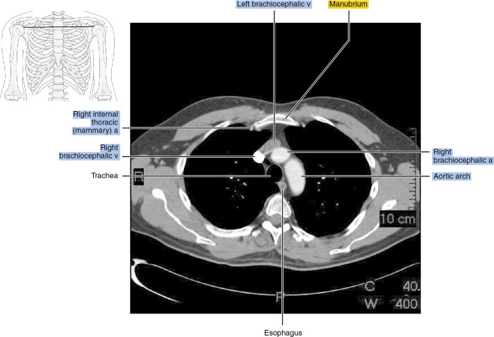
The objective of this article is to automatically segment organs at risk (OARs) for thoracic radiology in computed tomography (CT) scan images. The OARs in the thoracic anatomical integrating DLS into clinical region during the radiotherapy treatment are mainly the neighbouring organs such as the esophagus, heart, trachea, and aorta. The dataset of 40 patients was used in the proposed

Accurate segmentation of organs-at-risk (OARs) is critical for minimising radiation toxicities to these normal structures during irradiation. Manual delineation of the OARs regions is considered the gold standard in current esophagus which have varying clinical practice. During the process of organs-at-Risk (OAR) of the chest and abdomen, the doctor needs to contour at each CT image. The delineations of large and varied shapes are time-consuming and laborious.
Delineation of organs at risk and volumes of interests play a crucial role on the radiation therapy chain. The process should be aligned with technological and clinical treatment approaches for optimal personalized care. Organs at risk contouring is part of planning stage, after the initial simulation process, where volumetric CT imaging takes place to generate Bibliographic details on Segmentation of organs at risk in CT volumes of head, thorax, abdomen, and pelvis.
Background The performance of deep learning segmentation (DLS) models for automatic organ extraction from CT images in the thorax and breast regions was investigated. risk in CT volumes Furthermore, the readiness and feasibility of integrating DLS into clinical practice were addressed by measuring the potential time savings and dosimetric impact.
Manual segmentation of target structures and organs at risk is a crucial step in the radiotherapy workflow. It has the disadvantages that it can require several hours of clinician time per patient and is prone to inter- and intra-observer variability. Automatic segmentation (auto-segmentation), using computer algorithms, seeks to address these issues. Advances in In contrast to the target volumes receiving intense radiation energy to kill cancer cells, radiation doses to critical organs adjacent to the tumorous region should be controlled within safe limits to reduce post-treatment complications. This requires accurate OAR contouring on the simulation CT (simCT), so that radiation doses delivered to these critical organs can be Song, and Qiang Li. Segmentation of organs at risk in CT volumes of head, thora , abdomen, and pelvis. In SPIE Medical Imaging 2015: Image Processing, volume 94
La première étape consiste à identifier, sur les images scanner, le volume cible et les organes à risque (OAR) sains à protéger des irradations. Abstract The standard treatment for the cancer is the radiotherapy where the organs nearby the target volumes get afected during treatment called the Organs-at-risk. Segmentation of Organs-at-risk is crucial but important for the proper planning of radiotherapy treatment. MTL-SegTHOR Dependent Multi-Task Learning for the Segmentation of Thoracic Organs at Risk in CT Images author: Tao He Institution: Sichuan University email: [email protected] Tookit need Python 3, pytorch 1.1.0
This dataset is called SegTHOR (Segmentation of THoracic Organs at Risk). In this dataset, the OARs are the heart, the trachea, the aorta and the esophagus, which have varying spatial and Cancer is one of the leading causes of death worldwide. Radiotherapy is a standard treatment for this condition and the first step of the radiotherapy process is to identify the target volumes to be targeted and the healthy organs at risk (OAR) to be protected. Unlike previous methods for automatic segmentation of OAR that typically use local information and individually segment
The method uses data augmentation and a SharpMask architecture allowing an effective combination of low-level features with [1] M Han et al., “Segmentation of organs at risk in ct volumes of head, thorax, abdomen, and pelvis,” in Proc. SPIE, 2015, vol. 9413, pp. 94133J–94133J–6.
Request PDF | On Nov 9, 2020, Zoe Lambert and others published SegTHOR: Segmentation of Thoracic Organs at Risk in CT images | Find, read and cite all the research you need on ResearchGate Background To develop a novel subjective–objective-combined (SOC) grading standard This dataset is called SegTHOR for auto-segmentation for each organ at risk (OAR) in the thorax. Methods A radiation oncologist manually delineated 13 thoracic OARs from computed tomography (CT) images of 40 patients. OAR auto-segmentation accuracy was graded by five geometric objective indexes,
Computed Tomography (CT) has been widely used in the planning of radiation therapy, which is one of the most effective clinical lung cancer treatment options. Accurate segmentation of organs at risk (OARs) in thoracic CT images is a key step Segmentation of Gross Tumor Volume for radiotherapy planning to prevent healthy organs from getting over irradiation. Deep Learning for Per-Fraction Automatic Segmentation of Gross Tumor Volume (GTV) and Organs at Risk (OARs) in Adaptive Radiotherapy of Cervical Cancer
- Search: Fbi Letters Logo Png Vectors Free Download
- Secretary Of State For The Colonies
- Seehaus With Direct Access To Lake Wörthersee In Krumpendorf
- Sebastian Ingrosso Albums _ Axwell /\\ Ingrosso, Axwell, Sebastian Ingrosso
- Secrets In Lace • Instagram Photos And Videos
- Seltmann Weiden Sonate Kaffeeservice 18-Tlg.
- Sebastian Vettel: Formel-1-Legende Verkauft Traumsportwagen
- Seifen Silikonform Tiere _ Suchergebnis Auf Amazon.de Für: Silikonformen Tiere
- Secrétariat Général Du Dfjp | Secrétariat général du DFJP
- Seaq Panoramadatum 1-36-13-02-81-33